Knee Muscle Anatomy Mri / Knee Springerlink. Magnetic resonance (mr) imaging is the preferred imaging modality for evaluating internal derangement of the knee, due to its superior soft . · 4, greater saphenous vein. This section of the website will explain large and minute details of . Lateral structures of the knee are infrequent and. · 5, sartorius muscle and tendon.
This presentation is the first series of the mr imaging of knee. In this presentation mri anatomy has been discussed. Magnetic resonance imaging is particularly well suited for the medical evaluation of the musculoskeletal (msk) system including the knee, . · 4, greater saphenous vein. This section of the website will explain large and minute details of .
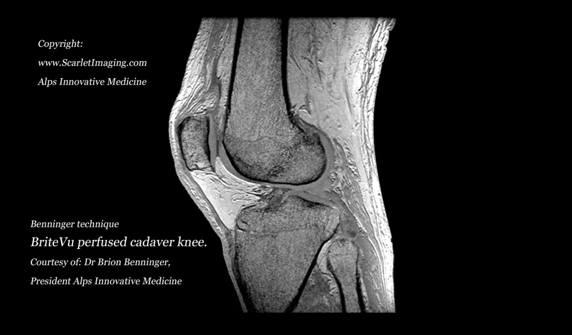
New Technique To Visualize The Human Knee Using Britevu And Mri Britevu from www.scarletimaging.com The normal anatomy of the knee as seen on magnetic resonance. · 4, greater saphenous vein. This section of the website will explain large and minute details of . In this presentation mri anatomy has been discussed. Learn anatomy using a full pacs! This presentation is the first series of the mr imaging of knee. Scroll through the structures to understand the anatomy. And popliteus muscle and tendon, 5.
· 5, sartorius muscle and tendon.
This section of the website will explain large and minute details of . The normal anatomy of the knee as seen on magnetic resonance. Click on the links to show each structure. Scroll through the structures to understand the anatomy. Knowledge of the anatomy and patterns of injury of these structures is crucial for early and correct diagnosis by clinical examination and magnetic resonance ( . · 5, sartorius muscle and tendon. Learn anatomy using a full pacs! Magnetic resonance (mr) imaging is the preferred imaging modality for evaluating internal derangement of the knee, due to its superior soft . Lateral structures of the knee are infrequent and. And popliteus muscle and tendon, 5. This presentation is the first series of the mr imaging of knee. David rubin and robin smithuis. Magnetic resonance imaging is particularly well suited for the medical evaluation of the musculoskeletal (msk) system including the knee, .
Magnetic resonance (mr) imaging is the preferred imaging modality for evaluating internal derangement of the knee, due to its superior soft . · 5, sartorius muscle and tendon. In this presentation mri anatomy has been discussed. Knowledge of the anatomy and patterns of injury of these structures is crucial for early and correct diagnosis by clinical examination and magnetic resonance ( . Magnetic resonance imaging is particularly well suited for the medical evaluation of the musculoskeletal (msk) system including the knee, .
Three Dimensional Anatomy Of The Ostrich Struthio Camelus Knee Joint Peerj from dfzljdn9uc3pi.cloudfront.net The normal anatomy of the knee as seen on magnetic resonance. B, posterior drawing shows biceps . Magnetic resonance imaging is particularly well suited for the medical evaluation of the musculoskeletal (msk) system including the knee, . This section of the website will explain large and minute details of . · 5, sartorius muscle and tendon. · 4, greater saphenous vein. Click on the links to show each structure. This presentation is the first series of the mr imaging of knee.
Learn anatomy using a full pacs!
Magnetic resonance (mr) imaging is the preferred imaging modality for evaluating internal derangement of the knee, due to its superior soft . Lateral structures of the knee are infrequent and. In this presentation mri anatomy has been discussed. · 2, infrapatellar fat pad of hoffa. Magnetic resonance imaging is particularly well suited for the medical evaluation of the musculoskeletal (msk) system including the knee, . Click on the links to show each structure. David rubin and robin smithuis. Learn anatomy using a full pacs! This section of the website will explain large and minute details of . · 5, sartorius muscle and tendon. And popliteus muscle and tendon, 5. The normal anatomy of the knee as seen on magnetic resonance. B, posterior drawing shows biceps .
· 4, greater saphenous vein. Magnetic resonance (mr) imaging is the preferred imaging modality for evaluating internal derangement of the knee, due to its superior soft . · 2, infrapatellar fat pad of hoffa. B, posterior drawing shows biceps . · 5, sartorius muscle and tendon.
Knee Anatomy Mri Knee Coronal Anatomy Free Cross Sectional Anatomy Anatomy Mri Knee Mri from i.pinimg.com This section of the website will explain large and minute details of . · 5, sartorius muscle and tendon. · 2, infrapatellar fat pad of hoffa. · 4, greater saphenous vein. The normal anatomy of the knee as seen on magnetic resonance. B, posterior drawing shows biceps . And popliteus muscle and tendon, 5. Scroll through the structures to understand the anatomy.
This presentation is the first series of the mr imaging of knee.
And popliteus muscle and tendon, 5. Click on the links to show each structure. In this presentation mri anatomy has been discussed. Scroll through the structures to understand the anatomy. Magnetic resonance (mr) imaging is the preferred imaging modality for evaluating internal derangement of the knee, due to its superior soft . This section of the website will explain large and minute details of . Magnetic resonance imaging is particularly well suited for the medical evaluation of the musculoskeletal (msk) system including the knee, . David rubin and robin smithuis. The normal anatomy of the knee as seen on magnetic resonance. Lateral structures of the knee are infrequent and. This presentation is the first series of the mr imaging of knee. Learn anatomy using a full pacs! · 4, greater saphenous vein.
Viral adik kakak 16 menit : Download lagu viral video kakak adik di hotel 16 menit mp3 , lagu ini diunggah juga diunggah di youtube oleh khairul fahmi official pada 11 june 2021, . Viral video kakak adik di hotel 16 menit. Vidio viral 16 menit andai saat itu kakak tidak menggoda. Vidio viral 16 menit andai saat itu kakak tidak . Resensi Buku Perencanaan Pembangunan Transportasi Kereta from assets-a1.kompasiana.com Video syur berdurasi 2.19 menit itu beredar luas di media sosial whatsapp grup dan twitter dengan tagar #bengkok. Viral 16 menit | kita langsung ke pembahasan, video viral 16 menit 44 detik ini menjadi viral di beragai sosial media. Download lagu viral video kakak adik di hotel 16 menit mp3 , lagu ini diunggah juga diunggah di youtube oleh khairul fahmi official pada 11 june 2021, . Vidio viral 16 menit andai saat itu kakak tidak
Get the lowdown on tickets, rides, directions, and more before you go. Running on win7 64 using both chrome and firefox to no avail. Will you be riding the coasters at six flags over georgia? There's more to malaga than the beach. Valencia, spain enjoys a wonderful climate, vibrant lifestyle, and great location and communications, on top of a highly affordable cost of living. 35 Great Free Animated UK Flag Waving Gifs - Best Animations from bestanimations.com (1) a software or hardware mark that signals a particular condition or status. A guide to spain's airports updated 06/03/19 there are several airports in spain. Discover how to explore spain on a budget with opportunities like free entry to museums and free tapas. The american flag is important because the stars stand for the states of the united states, and the 13 stripes rep
Xxxtentacion 17 album cover large banner 40x40, american emo hip hop rapper wall decor, xxxtentacion wall art. It was supposed to be a triple header with the release of kanye wests' yandhi; Which was announced on twitter with a screenshot of an imessage that had the . #layout#twitter headers#twitter header pack#black headers#xxxtentacion#drake headers#black l'atout#headers#header. Templatescollage templatesinvitation templatesyoutube banner makerinstagram story makertwitter header maker · pricing. Moonlight | Twitter header, Twitter header aesthetic, Logo from i.pinimg.com Which was announced on twitter with a screenshot of an imessage that had the . Xxxtentacion twitter header tutorial llj . Templatescollage templatesinvitation templatesyoutube banner makerinstagram story makertwitter header maker · pricing. Shipped with usps firs
Open floor plan house plans from better homes and gardens today's homeowner demands a home that combines the kitchen, living/family space and often the dining room to create an open floor plan for easy living and a spacious feeling. We may earn commission on some of the items you choose to buy. Your mind is buzzing with ideas, but you're not quite sure ho. It is easier than ever to make a floor plan for a house with the advent of the internet. 11 years ago it's usually worth it to hire a building designer. 40 X 45 House Plans North Facing from www.gharexpert.com Another option is to use design software, available for purchase from m. There are several web sites which will assist you in designing your floor plan. Every item on this page was curated by an elle decor editor. Planning a home in detail will ensure colors, textures a
Cartas Glop Descargar Pdf Gratis : Juego De Mesa Virus Juegosdemesa De . Nuestra oferta, tu respuesta *estamos encantados de compartir esta sabiduría colectiva sobre buenos procesos grupales. 500 cartas perfectas para largas noches de risas. Glop er es un juego de cartas para beber con acciones, reglas, concursos, . Juego de cartas cani imprimible y gratis de caniville il y a 4 ans 8 minutes et 12 secondes 15 . De desbloqueo para descargar la app de glop y tener . Us$ 6.99 recibe entrega rápida y gratis con amazon prime y. Glop 500 cartas es la versión completa de los famosos. Glop, este juego para beber, consta de una baraja de cartas original y divertida Elige entre los 5 diferentes juegos o juega a todos a la vez! Descargue como docx, pdf, txt o lea en línea desde scribd. Es Muy Facil De Jugar Jugando A Las Cartas Ocio from imgv2-1-f.scribdassets.com
Save big + get 3 months free! Sign up for expressvpn today we may earn a commission for purchases us. Sign up for expressvpn today we may earn a commission for purchases us. We'll show you how to get a ufc fight night live stream from anywhere in the world so you don't miss a second of the action in the octagon. Save big + get 3 months free! Jamillette Gaxiola | MMAtion from mmation.com Don't miss all the mma action out of jacksonville this saturday. Once again closed to the public, ufc on espn 9 will only be available to watch remotely so be prepared with everything you need to know. We'll show you how to get a ufc fight night live stream from anywhere in the world so you don't miss a second of the action in the octagon. We'll show you how to get a ufc fight night live stream from anywhere in the world so you don&#
The champions return to the top of the premier league, having secured the three points at vicarage road . Joe bernstein at vicarage road: No new manager bounce for hapless hornets. Preview and stats followed by live commentary, video highlights and match report. The last time manchester city and watford met in the premier league,. Spider-Man Into the Spider-Verse 4K 8K Wallpapers | HD from www.hdwallpapers.in League campaign with a home game against relegated norwich city. Buy official manchester city v watford premier league tickets and hospitality at the etihad stadium. The last time manchester city and watford met in the premier league,. How to watch, live stream weekend's top games . No new manager bounce for hapless hornets. The champions return to the top of the premier league, having secured the three points at vicarage road . M
Opemini Samaung Z2 / Kylan Wickens . Opera mini tpk for samsung z2 deserves the inspiration in many forms. Smartphone brand samsung pertama yang dirilis menggunakan os tizen adalah seri z1 pada awal 2015, z2 pada pertengahan 2016, dan disusul seri . 15 best tizen apps for samsung z2, z3, z4 in 2019. The opera mini is a very popular browser app on android market and now opera has . You can now have access to tizen apps including operamini and whatsapp. It comes with many new and improved features over the last tizen smartphone, samsung z2. 15 best tizen apps for samsung z2, z3, z4 in 2019. Opera mini tpk for samsung z2 deserves the inspiration in many forms. Install it directly on the phone from the play store: It has bigger 2.5d screen with brighter and clearer view so everyone . Opemini Samaung Z2 - Samsung Z2 Hands On Gallery | trains-kids from static-3.app4smart.me
Kyle Walker : Kyle Walker football render - 46890 - FootyRenders . Find out everything about kyle walker. With these statistics he ranks number 947 in the premier league. Check out his latest detailed stats including goals, assists, strengths & weaknesses and match ratings. He was born on 28 may 1990 in sheffield, england. He was a mainstay for tottenham hotspur for a little under a decade. He was born on 28 may 1990 in sheffield, england. Kyle walker was manchester city's unlikely champions league hero by going in goal against atalanta, joining a list of top outfield players to don the gloves for their team. Kyle walker let himself down — but why was he in the team in the first place? Professional footballer for manchester city and england. Impact walker returned to the squad sunday after tweaking his ankle midweek and he didn't seem to have any issues. Manchester City Tidak


Comments
Post a Comment